
| NeuroAids Vol. 3, Issue 5 (October 2000) |
A Rapid, Reproducible Model of AIDS and Encephalitis in SIV infected Macaques Demonstrates the Role of Viral Load in CNS DiseaseM. C. Zink1 and J. E. Clements1 E-mail: mczink@jhmi.edu , jclement@jhmi.edu |
| Abstract |
|---|
| Abstract | Introduction | A Rapid SIV/Macaque Model of AIDS Dementia | High CSF Viral Load and SIV Encephalitis | Conclusions | References | ||||
| The development of HIV-1 associated dementia usually occurs in later stages of AIDS in approximately 25% of HIV-infected individuals. In the SIV animal model for AIDS, CNS disease also occurs in the later stages of disease, 2-3 years after infection and in only a small percentage of the infected macaques. In order to examine the mechanisms involved in the development of CNS disease in the SIV model, a model was need in which a high proportion of the macaques developed CNS lesions in a relatively short period of time. Macaques were infected with a neurovirulent virus, SIV/17E-Fr, and an immunosuppressive virus swarm, SIV/deltaB670 and these animals developed AIDS within 12 weeks and a high proportion had CNS lesions. The role of virus load in the plasma, cerebrospinal fluid (CSF) and brain were examined in this model and high viral load in CSF was found to correlate with the severity of CNS lesions and SIV encephalitis. Further, the presence of viral RNA in macaque brain correlated with the presence of CNS lesions and SIV encephalitis. Animals that had little or no signs of SIV encephalitis had no detectable viral RNA |
| Introduction |
|---|
| Abstract | Introduction | A Rapid SIV/Macaque Model of AIDS Dementia | High CSF Viral Load and SIV Encephalitis | Conclusions | References | ||||
|
HIV-1 associated dementia affects approximately 25% of HIV-infected individuals, and pathological changes are seen in the brains of 70 to 90% of HIV-infected people (1)(2)(3). Most individuals with HIV-associated dementia are in the terminal, immunosuppressive, stages of disease, suggesting that intact immune responses may protect the CNS from the alterations that result in dementia (4). SIV infection of macaques has been shown to recapitulate key features of HIV infection of the human CNS, including the development of encephalitis with characteristic histopathological changes, psychomotor impairment and sensory evoked potentials (5)(6)(7). However, in most SIV models and similar to HIV infection in people, both the incidence and time to onset of CNS disease are variable. In SIV infection of macaques, typically 30% of macaques develop encephalitis, and the time to development of CNS disease is 1 to 3 years. The need for large numbers of animals and the long periods required for the development of disease thus greatly complicates the design and interpretation of studies. |
| A Rapid SIV/Macaque Model of AIDS Dementia |
|---|
| Abstract | Introduction | A Rapid SIV/Macaque Model of AIDS Dementia | High CSF Viral Load and SIV Encephalitis | Conclusions | References | ||||
|
We hypothesized that the delay between inoculation and the development of CNS disease was related to both viral and host factors, and that by manipulating these factors, we might be able to increase the percentage of animals that develop CNS lesions and shorten the period of clinical latency. Our model of rapid development of CNS disease in a large proportion of animals was predicated on three specific observations. First, SIV strains vary greatly in the degree to which they cause neurological disease. Many SIV strains used in macaque infection studies are only modestly neurovirulent, causing neurological disease in less than half of inoculated animals. Second, most HIV-infected individuals develop neurological disease concurrent with the development of terminal immunosuppression. Third, our previous studies comparing the severity of neurological lesions in rhesus macaques, pigtailed macaques, and cynomolgus macaques suggested that pigtailed macaques may be more sensitive to the development of neurological lesions than the other two species (6). We therefore developed a model using pigtailed macaques that involved the co-inoculation of a highly neurovirulent, macrophage-tropic virus, SIV/17E-Fr (8)(9)(10) and a highly immunosuppressive virus swarm of SIV, SIV/deltaB670 (11) that also contained neurovirulent virus. Our intention was to expose the macaque species most susceptible to the development of CNS lesions to a reliably neurovirulent virus so that the virus would become established in the brain prior to the development of antiviral immune responses. Although replication of this virus in the brain is probably controlled in response to the acute immune responses, virus replication would increase with the loss of immune control upon the development of immunosuppression. Twelve pigtailed macaques were inoculated with the dual virus inoculum and euthanized at 3 months postinoculation, 11 (92%) have developed SIV encephalitis, with the CNS lesions classified as severe in 4 macaques, moderate in 5, and mild in 2 (6). Macaques infected with this combination of viruses develop profound and rapid declines in CD4+ T cell counts in peripheral blood (12). This is accompanied by rapid increases and sustained high levels of viral RNA in the peripheral blood (12). This pattern of rapidly developing immunosuppression and high levels of plasma viral load is similar to findings in HIV-infected individuals who are termed rapid progressors. This model provides an important tool in understanding the pathogenesis of HIV dementia in humans. It allows the dissection of virological, immunological and pathological events occurring in both the brain and the periphery at various stages of infection, particularly, acute infection that is especially difficult to study in humans infected with HIV. We have already used this model to determine whether viral load in the plasma and/or CSF at various stages of infection correlate with the development of neurological disease (see Topic 2 below). Studies are currently under way to determine the role of various pro-inflammatory cytokines and chemokines in the development of inflammatory changes and neuronal loss.
|
|
High
CSF Viral Load and SIV Encephalitis
|
|---|
| Abstract | Introduction | A Rapid SIV/Macaque Model of AIDS Dementia | High CSF Viral Load and SIV Encephalitis | Conclusions | References | ||||
|
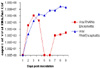
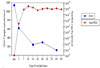
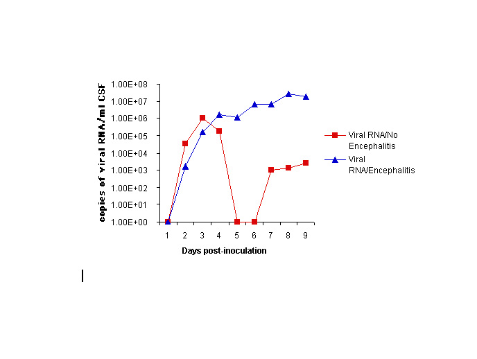
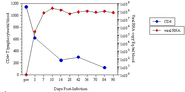
This rapid model of SIV encephalitis has provided new insights into the pathogenesis of SIV encephalitis. In one study, using 6 macaques we examined the relationship between viral load in the CSF throughout infection and the development and severity of neurological lesions (12). Viral RNA was measured by real-time RT-PCR in blood and CSF obtained at 7, 10, 14, and 28 days post-inoculation and every two weeks thereafter until 3 months post-inoculation when the macaques were euthanized. Viral RNA in plasma rose in the first two weeks of infection and stayed at high levels between 107 and 108 throughout the infection (Figure 1). However, all infected macaques had high plasma viral load and there was no correlation between the level of viral load in the plasma and the development of SIV encephalitis. Viral RNA in CSF rose to between 104 to 106 during acute infection in all 6 inoculated macaques, peaking between 10 and 14 days post-inoculation (Figure 2). After 14 days p.i., viral RNA levels in the 5 macaques that eventually developed SIV encephalitis stayed stable or continued to rise. In contrast, CSF viral load dropped to below detectable levels by 28 days p.i. in the single macaque that did not develop CNS lesions. This study indicated that the level of viral RNA in CSF after acute infection is predictive of the development of neurological lesions and demonstrated the utility of the rapid dual inoculation model.
Viral load in the four regions of the brain (basal ganglia, thalamus, parietal cortex and cerebellum) was determined in all six macaques. The macaque that did not develop CNS lesions had no detectable viral RNA in any of the regions of the brain. In contrast, all five of the animals with CNS lesions had significant levels of RNA in the brain (from 106 to 108 copies of viral RNA/ug total RNA) (12). We have now examined a total of 30 dual-inoculated macaques euthanized during the acute (10 days p.i.), post-acute (21 days p.i.), pre-terminal (8 weeks p.i.) and terminal (3 months) stages of infection. The model has provided novel insights into the role of active virus replication in the CNS and the development of lesions, the CNS as a reservoir for minimally replicating virus and a variety of biochemical, virological, and immunological changes that occur in the brain and other organs during infection. Such studies should provide a mechanistic understanding of the events that initiate and perpetuate inflammation in the CNS and the critical events that lead to AIDS dementia. |
| Conclusions |
|---|
| Abstract | Introduction | A Rapid SIV/Macaques Model of AIDS Dementia | High CSF Viral Load and SIV Encephalitis | Conclusions | References | ||||
|
The SIV model provides an excellent model to study the pathogenesis of AIDS and AIDS dementia. The development of the rapid, reproducible SIV model of AIDS and CNS disease provides an improved system to examine the development of CNS disease. In particular, this model is being used to identify viral and cellular markers that are prognostic for the development of CNS disease. Because this model is highly reproducible, there is a great deal of statistical power in animal groups of 6 and statistically significant results have been obtained with each of the animal groups. Thus, this model will also be valuable to test emerging drugs developed specifically for CNS infection and disease. |
| Abstract | Introduction | A Rapid SIV/Macaques Model of AIDS Dementia | High CSF Viral Load and SIV Encephalitis | Conclusions | References | ||||
|
Use the Back button in your browser to continue reading the article.
(2) Spencer DC, Price RW (1992). Human immunodeficiency virus and the central nervous system. Annu.Rev.Microbiol. 46, 655-693. Medline (3) McArthur J, Hoover D, et al. (1993). Dementia in AIDS patients: incidence and risk factors. Multicenter AIDS Cohort Study. Neurology Nov 43(11), 2245-2253. Medline (4) Soontornniyomkij V, Nieto-Rodriguez JA, et al. (1998). Brain HIV burden and length of survival after AIDS diagnosis. Clin Neuropathol. Mar-Apr 17(2), 95-9. Medline (5) Murphy PM (1996). Chemokine receptors: structure, function and role in microbial pathogenesis. Cytokine Growth Factor Reviews. June 7(1), 47-64.Medline (6) Zink MC, Amedee AM, et al. (1997). Pathogenesis of SIV encephalitis. Selection and replication of neurovirulent SIV. Am. J. Pathol. Sep 151(3), 793-803. Medline (7) Raymond LA, Wallace D, et al. (2000). Sensory evoked potentials in SIV-infected monkeys with rapidly and slowly progressing disease [In Process Citation]. AIDS Res Hum Retroviruses Aug 10 16(12), 1163-73. Medline (8) Mankowski JL, Spelman JP et al. (1994). Neurovirulent simian immunodeficiency virus replicates productively in endothelial cells of the central nervous system in vivo and in vitro. J Virol Dec 68(12), 8202-8. Medline (9) Flaherty MT, Hauer DA, et al. (1997). Molecular and biological characterization of a neurovirulent molecular clone of SIV. J.Virol. Aug 71(8), 5790-5798. Medline (10) Mankowski JL, Flaherty MT, et al. (1997). Pathogenesis of SIV encephalitis: Viral determinants of neurovirulence. J.Virol. Aug 71(8), 6055-6060. Medline (11) Sharer LR, Michaels J, et al. (1991). Serial pathogenesis study of SIV brain infection. J.Med.Primatol. June 20(4), 211-217. Medline (12) Zink MC, Suryanarayana K, et al. (1999). High viral load in CSF and brain correlates with severity of SIV encephalitis. J.Virol. Dec 73(12), 10480-8. Medline |
|
NeuroAids is a project of Science OnLine funded through a grant from the National Institute of Mental Health. |
|

|
Copyright ©1998 by AAAS Science Publications, Inc. |