| NeuroAids Vol. 3, Issue 2 (March 2000) |
The Blood-Brain Barrier in HIV-associated Dementia C.F. Pereira1, H.S.L.M. Nottet1 E-mail: h.s.l.m.nottet@lab.azu.nl |
| Abstract |
|---|
| Abstract | Introduction | Transmigration
through BBB |
Transmigration
through brain parenchym |
Conclusions | References | ||||
|
Monocytes have been shown to infiltrate brain tissue during various neurological disorders including HIV-associated dementia. The presence of an excess of activated macrophages in brain tissue is accompanied by a loss in neuronal function and viability. Therapeutic options against such neurological disorders could be aimed at the prevention of monocyte infiltration across the blood-brain barrier. Therefore, a better understanding of these processes is needed. Recent insights in cellular processes between monocytes/macrophages and brain microvascular endothelial cells in the neuropathogenesis of HIV-1 infection demonstrate that monocytes roll along endothelial cells via the inducible endothelial adhesion molecule E-selectin. Binding of these cells is mainly mediated via the endothelial adhesion molecule named vascular cell adhesion molecule-1. The transmigration through the blood-brain barrier is facilitated by both endothelial and monocyte/macrophage-derived nitric oxide, as well as by the increased production of gelatinase B activity by HIV-infected monocytes/macrophages. Chemokines produced within the brain regulate the traffic of infiltrating monocytes through the brain parenchyma. In addition, endothelial cells also produce monocyte-attracting chemokines during their interactions with HIV-infected monocytes/macrophages, thus promoting additional influx of mononuclear phagocytes into the brain. Furthermore, excessive infiltration of monocytes is accompanied by endothelial damage resulting in the loss of tight junctions. In summary, brain microvascular endothelial cells might contribute to the neuropathogenesis of HIV-1 infection. |
| Introduction |
|---|
| Abstract | Introduction | Transmigration
through BBB |
Transmigration
through brain parenchym |
Conclusions | References | ||||
|
HIV-associated dementia (HAD) is the most severe neurological manifestation of HIV infection of the brain. In the era before treatment with anti-HIV compounds 30% of the adults with AIDS and 50% of the pediatric AIDS cases were affected by HIV-induced neurological complications in the western world. Interestingly, in the adult brain HIV only productively replicates in macrophages/microglia and not in neurons, astrocytes, oligodendrocytes or brain microvascular endothelial cells (BMEC) (1)(2)(3). Neuropathological features of HIV-1 encephalitis include productive HIV-1 infection of brain cells of monocyte/macrophage lineage, multinucleated giant cell formation, monocyte infiltration of the central nervous system (CNS), astrogliosis, and myelin pallor. Some neurons undergo dendritic pruning, simplification of synaptic contacts, and frank cell loss (4). These neuronal changes are clinically manifested by cognitive and motor dysfunctions that affect many individuals with AIDS. The finding that neurological symptoms are a consequence of dendritic injury and synaptic loss (5), and occur in absence of neuronal cell loss (6), suggest that treatment may retard the onset of cognitive and motor defects. HIV- l is a haematogenously spread virus that most likely gains entry into the brain inside blood-derived macrophages (7). Indeed, HIV-1 is selectively localized within perivascular and infiltrated parenchymal blood-derived macrophages and microglia (1)(2)(3). Axonal spread of HIV as a mechanism to enter the CNS is ruled out since direct neuronal infection has not been demonstrated in pathologic specimens of AIDS patients with HIV-1 encephalitis. Theoretically, cell-free HIV might infect BMEC and subsequent release of virus at the abluminal membrane of the blood-brain barrier (BBB) would result in HIV entry into the brain parenchyma. Although direct in vitro infection of brain endothelium by lymphotropic HIV-l has been described, the relevance of that finding remains questionable (8). First, macrophagetropic HIV-1 is the predominant viral phenotype isolated during the acute seroconversion reaction, thus excluding a role for lymphotropic HIV-1 in CNS infiltration (9). Second, brain-derived viral strains are macrophagetropic rather than lymphotropic (10)(11). Third, there is no in vivo evidence for HIV infection of brain endothelial cells (1)(2)(3). In addition, several other investigators could not find in vitro evidence for permissiveness of these cells to either lymphotropic or macrophagetropic HIV-1 (12)(13)(14). These findings and the ample evidence that only blood-derived macrophages and microglia support productive viral replication within the CNS suggest that HIV-1 gains entry into the brain within macrophages or as as cell-free virions (15). Although the early transmigration of HIV-infected monocytes/macrophages (M/M) into the CNS might result in meningitis, the occurrence of severe neurological symptoms including dementia take place at a relatively late stage of HIV infection. In these stages the immune system is deteriorated and activated. This has consequences for the mechanisms involved in the brain infiltration by HIV-infected M/M and the subsequent development of HAD as outlined hereafter. |
| Transmigration of HIV-infected M/M through the blood-brain barrier |
|---|
| Abstract | Introduction | Transmigration
through BBB |
Transmigration
through brain parenchym |
Conclusions | References | ||||
|
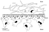
During the later symptomatic stages of HIV-1 infection elevated levels of pro-inflammatory cytokines such as tumor necrosis factor-a (TNF-a) and interleukin-1b (IL-1b) have been detected in serum of AIDS patients and in macrophages isolated from HIV-infected individuals (16)(17). Both cytokines are mediators of meningitis and regulate BBB permeability in a reversible manner. Indeed, intracisternal injection of TNF-a and IL-1b into rats increased BBB permeability for systemically administrated radioactive tracers (18). In addition, these cytokines induce the expression of endothelial adhesion molecules that facilitate rolling and binding of leukocytes. Indeed, activated HIV-infected M/M induce the highest levels of E-selectin and vascular cell adhesion molecule-1 (VCAM-1) on brain microvascular endothelial cells (14) as compared to non-activated HIV-infected M/M and activated uninfected M/M (Figure 1). Since E-selectin mediates rolling and VCAM-1 mediates binding of leukocytes to endothelium, HIV-infected cells have an advantage in these processes when compared to normal M/M. The in vitro studies are supported by the observation that endothelial expression of E-selectin and to a lesser degree VCAM-1 was the highest in brain tissue of demented AIDS patients when compared to non-demented AIDS patients (14). In addition, monocyte binding to encephalitic brain could be prevented by blocking monoclonal antibodies directed against both adhesion molecules (14). Since recombinant tat induces E-selectin expression on other endothelial cells, the induction of this adhesion molecule is most likely mediated by the HIV-1 tat molecule that is secreted by HIV-infected M/M (19). Furthermore, HIV-1 infection of M/M induces the production of low levels of nitric oxide (NO), a potent vasoactive molecule (20)(21). Since NO can slow down the speed of blood cells passing through the blood vessel, the production of NO might increase the change that mononuclear cells roll on endothelium via E-selectin (Figure 1).
However, besides these subtle changes between M/M and endothelial cells there is also evidence of endothelial damage that might underlie the excessive monocyte infiltration into the brain and even contribute to the severe stages of HAD. For instance, in vitro HIV-1 infection of M/M resulted in superoxide anion production (21), and endothelial NO synthase was induced in cocultures of M/M and endothelial cells (unpublished data). Since superoxide anion and NO form the highly toxic molecule peroxynitrite, immunohistochemical staining for nitrotyrosine, the footprint of peroxynitrite, was performed and showed extensive immunoreactivity in perivascular areas of brain tissue of demented AIDS patients (21). Furthermore, the architecture of many bloodvessels was damaged as shown by immunohistochemical staining for zonula occludens-1, a tight junction protein (23). Thus, during the intimate contact with HIV-infected M/M endothelial cells may contribute to their own damage by the production of NO. These findings might explain the increased permeability of artificial BBB for radioactive albumin when put in contact with HIV-infected M/M (24). After their migration through the endothelial cell layer, the monocytes will encounter the basement membrane that surrounds the abluminal side of the BBB. HIV-infected M/M produce higher levels of gelatinase B as compared to uninfected M/M. These cells therefore have an advantage in the process of diapedesis since gelatinase B degrades the extracellular matrix proteins that underlie the endothelial cell monolayer and in such a way increases the permeability of the BBB (25). Interestingly, incubation of HIV-infected M/M with tissue inhibitor of metalloproteinase-1 (TIMP-1) and TIMP-2 partially inhibited the increased permeability of endothelial cell monolayers to 125I albumin. These proteinase inhibitors antagonize the effects of matrix metalloproteinases, such as the HIV-induced gelatinase B activity (25). Importantly, matrix metalloproteinase-9 activity in the cerebrospinal fluid of HIV-infected patients was significantly higher in patients with neurological deficits than in patients without neurological deficits (26). This finding suggest that, in addition to the effects of HIV-infected M/M on endothelial cells, interactions between HIV-infected M/M and the extracellular matrix also affect BBB permeability (Figure 1). Indeed, cocultivation of HIV-infected M/M with BMEC resulted in an increased permeability of the endothelium, measured as 125I albumin passage through endothelial cell monolayers in transwell systems (24). In addition, immune-activated M/M placed on BMEC cultured on the upper chamber of a transwell migrated in larger numbers through the artificial BBB than non-activated cells (27). |
| Migration of HIV-infected M/M through the brain parenchyma |
|---|
| Abstract | Introduction | Transmigration
through BBB |
Transmigration
through brain parenchym |
Conclusions | References | ||||
|
The distribution of infected macrophages in the brain demonstrates viral predisposition for cerebral white matter, deep gray matter (basal ganglia and thalamus), and the brain-stem. Although the exact mechanisms that underlie this distribution remain unknown, temporal expression of adhesion molecules and chemokines in the brain has been suggested to guide specific trafficking of HIV-infected M/M to these brain areas (28). In any event, chemoattractants have been detected in brain tissue of AIDS patients with HIV- l encephalitis, and might therefore play a role both in infiltration and subsequent migration of HIV-infected M/M into the brain (Figure 1). For instance, transforming growth factor-b (TGF-b), a cytokine that induces the migration of monocytes at femtomolar concentrations, was clearly identified within AIDS brain tissues in association with macrophages and astrocytes, but not in control brain tissue (29). More recently, elevated expression of the b chemokines macrophage inflammatory protein- (MIP-1a) and MIP-1b was detected in brain tissue of demented AIDS patients as compared to non-demented AIDS patients (30)(31). Cells expressing these chemokines were identified morphologically as brain macrophages and astrocytes. Interestingly, MIP-1b levels were much more prevalent than the MIP-la levels (30)(31). Since the monocyte chemotactic activity of MIP-la is much more potent than that of MIP-1b, the elevated levels of MIP-1a in HIV-infected brain tissue might result in further monocyte infiltration of brain tissue and subsequent spread of viral CNS infection. In addition, monocyte chemoattractant protein-1 (MCP-1), another chemokine with relatively selective chemoattractant properties for monocytes, was found to be elevated in astrocytes in brain tissue of demented AIDS patients (31)(32). Furthermore, elevated levels of MCP-1 in CSF of demented individuals have also been described (32). Indeed, using a coculture of human endothelial cells and astrocytes to model the BBB, it was recently demonstrated that tat-induced, astrocyte-derived MCP-1 directs the transmigration of monocytes (33). And, finally, microglial expression of RANTES (Regulated upon Activation, Normal T-cell Expressed and Secreted) was found to be elevated in brain tissue of demented AIDS patients as compared to non-demented AIDS patients (31). Interestingly, endothelial cells themselves might also play a role in the recruitment of monocytes into the CNS by the production of chemokines. For instance, these cells elicit the production of several chemokines by uninfected as well as HIV-infected M/M (22). The levels produced by the HIV-infected cocultures are significantly higher than the levels produced by the uninfected cocultures. Importantly, endothelial expression of MCP-1 was readily detected in brain tissue of AIDS patients with HAD when compared to tissue of non-demented AIDS patients (22). |
| Conclusions |
|---|
| Abstract | Introduction | Transmigration
through BBB |
Transmigration
through brain parenchym |
Conclusions | References | ||||
|
HIV-1 enters the brain in a haematogenously fashion early during HIV-1 infection. Experimental evidence has been provided by several scientific investigators that it gains entry via the Trojan horse model by modulating both monocyte/macrophage and endothelial functions. HIV takes advantage of cellular machineries involved in rolling and binding processes between both cells and also increases the ability for monocytes to transmigrate into the CNS. Apparently the brain is able to limit extensive HIV-1 replication within the CNS or is able to suppress inflammatory processes. However, when the immune system is deteriorated, neurological complications start to occur and will eventually lead to dementia. During these events circulating monocytes and possibly also macrophages resident within the brain are immune-activated leading to enhanced production of molecules that increase BBB permeability, endothelial adhesion molecules expression, and chemokine expression. Eventually, when there is an excess of infiltrating monocytes into the brain the BBB is damaged with concomitant loss of tight junctions. At these stages, the BMEC might, in addition to the HIV-infected monocyte/macrophages, also function as instigators of the BBB loss and also contribute to the neurological complications. Acknowledgments |
| Abstract | Introduction | Transmigration
through BBB |
Transmigration
through brain parenchym |
Conclusions | References | ||||
|
Use the Back button in your browser to continue reading the article. (1) Koenig S, Gendelman HE, Orenstein JM, Dal Canto MC, Pezeshkpour GH, Yungbluth M, Janotta F, Aksamit A, Martin MA, Fauci AS (1986). Detection of AIDS virus in macrophages in brain tissue from AIDS patients with encephalopathy. Science 233: 1089-93. Medline (2) Wiley CA, Schrier RD, Nelson JA, Lampert PW, Oldstone MB (1986). Cellular localization of human immunodeficiency virus infection within the brains of acquired immune deficiency syndrome patients. Proc. Natl. Acad. Sci. USA 83: 7089-93. Medline (3) Vazeux R, Brousse N, Jarry A, Henin D, Marche C, Vedrenne C, Mikol J, Wolff M, Michon C, Rozenbaum W, Bureau JF, Montagnier L, Brahic M (1987). AIDS subacute encephalitis. Identification of HIV-infected cells. Am. J. Pathol. 126: 403-10. Medline (4) Masliah E, Achim CL, Ge N, DeTeresa R, Terry RD, Wiley CA (1992). Spectrum of human immunodeficiency virus-associated neocortical damage. Ann. Neurol. 32: 321-9. Medline (5) Masliah E, Heaton RK, Marcotte TD, Ellis RJ, Wiley CA, Mallory M, Achim CL, McCutchan JA, Nelson JA, Atkinson JH, Grant I, the HNCR Group (1997). Dendritic injury is a pathological substrate for human immunodeficiency virus-related cognitive disorders. Ann. Neurol. 42: 963-72. Medline (6) Seilhean D, Duyckaerts C, Vazeux R, Bolgert F, Brunet P, Katlama C, Gentilini M, Hauw JJ (1993). HIV-1-associated cognitive/motor complex: absence of neuronal loss in the cerebral neocortex. Neurology 43: 1492-9. Medline (7) Nottet HSLM, Gendelman HE (1995). Unraveling the neuroimmune mechanisms for the HIV-1-associated cognitive/motor complex. Immunol. Today 16: 441-8. Medline (8) Moses AV, Bloom FE, Pauza CD, Nelson JA (1993). HIV infection of human brain capillary endothelial cells occurs via a CD4/galactosylceramide-independent mechanism. Proc Natl Acad Sci USA 90: 10474-8. Medline (9) Zhu T, Mo H, Wang N, Nam DS, Cao Y, Koup RA, Ho DD (1993). Genotypic and phenotypic characterisation of HIV-1 in patients with primary infection. Science 261: 1179-81. Medline (10) Sharpless NE, O'Brien WA, Verdin E, Kufta CV, Chen ISY, Dubois-Dalcq M (1992). Human immunodeficiency virus type l tropism for brain microglial cells is determined by a region of the env glycoprotein that controls macrophage tropism. J. Virol. 66: 2588-93. Medline (11) Korber BTM, Kunstman KJ, Patterson BK, Furtado M, McEvilly MM, Levy R, Wolinsky SM (1994). Genetic differences between blood- and brain-derived viral sequences from human immunodeficiency virus type l-infected patients: Evidence of conserved elements in the V3 region of the envelope protein of brain-derived sequences. J. Virol. 68: 7467-81. Medline (12) Gilles PN, Lathey JL, Spector SA (1995). Replication of macrophagetropic and T-cell-tropic strains of human immunodeficiency virus type l is augmented by macrophage-endothelial contact. J. Virol. 69: 2133-9. Medline (13) Poland SD, Rice GPA, Dekaban GA (1995). HIV-1 infection of human brain-derived microvascular endothelial cells in vitro. J. Acquir. Immune Defic. Syndr. Hum. Retrov. 8: 437-45. Medline (14) Nottet HSLM, Persidsky Y, Sasseville VG, Nakuna AN, Bock P, Zhai Q-H, Sharer LR, McComb RD, Swindels S, Soderland C, Gendelman HE (1996). Mechanisms for the transendothelial migration of HIV-1 infected monocytes into brain. J. Immunol. 156: 1284-95. Medline (15) Nottet HSLM (1999). Interactions between macrophages and brain microvascular endothelial cells: role in pathogenesis of HIV-1 infection and blood-brain barrier function. J. Neurovirol. 5: 659-69. Medline (16) Lahdevirta J, Maury CPJ, Teppo AM, Repo H (1988). Elevated levels of circulating eachectin/tumor necrosis factor in patients with acquired immunodeficiency syndrome. Am. J. Med. 85: 289-91. Medline (17) Buhl R, Jaffe H, Holroyd K, Borok Z, Roum JH, Mastrangeli A, Wells FB, Kirby M, Saltini C, Crystal RG (1993). Activation of alveolar macrophages in asymptomatic HIV-infected individuals. J. Immunol. 150: 1019-28. Medline (18) Quagliarello VJ, Wispelwey B, Long WJ Jr. Scheld WM (1991). Recombinant human interleukin-1 induces meningitis and blood-brain barrier injury in the rat. Characterisation and comparison with tumor necrosis factor. J. Clin.Invest. 87:1360-6. Medline (19) Hofman FM, Wright AD, Dohadwala MM, Wong-Staal F, Walker SM (1993). Exogenous Tat protein activates human endothelial cells. Blood 92: 2774-80. Medline (20) Bukrinsky MI, Nottet HSLM, Schmidtmayerova H, Dubrovsky L, Flanagan CR, Mullins ME, Lipton SA, Gendelman HE (1995). Regulation of nitric oxide synthase activity in human immunodeficiency virus type 1 (HIV-1)-infected monocytes: implications for HIV-associated neurological disease. J. Exp. Med. 181: 735-45. Medline (21) Boven LA, Gomes L, Hery C, Gray F, Verhoef J, Portegies P, Tardieu M, Nottet HSLM (1999). Increased peroxynitrite activity in AIDS Dementia Complex: implications for the neuropathogenesis of HIV-1 infection. J. Immunol. 162: 4319-27. Medline (22) Boven LA, Brey E, Middel J, Schotte D, Verhoef J, Soderland C, Nottet HSLM. Interactions between HIV-infected monocyte-derived macrophages and brain microvascular endothelial cells result in increased production of CC chemokines. J. Neurovirol. 2000; in press. (23) Boven LA, Middel J, Verhoef J, de Groot CJA, Nottet HSLM. Macrophage infiltration is highly associated with loss of the tight junction protein zonula occludens in AIDS dementia complex. Neuropath. Appl. Neuro. 2000; in press. (24) Dhawan S, Toro LA, Jones E, Meltzer MS (1992). Interactions between HIV-infected monocytes and the extracellular matrix: HIV-infected monocytes secrete neutral metalloproteases that degrade basement membrane protein matrices. J. Leukoc. Biol. 52: 244-8. Medline (25) Dhawan S, Weeks BS, Soderland C, Schnaper HW, Toro LA, Asthana SP, Hewlett IK, Stetler-Stevenson WG, Yamada SS, Yamada KM, Meltzer MS (1995). HIV-1 infection alters monocyte interactions with human microvascular endothelial cells. J. Immunol. 154: 422-32. Medline (26) Sporer B, Paul R, Koedel U, Grimm R, Wick M, Goebel FD, Pfister H (1998). Presence of matrix metalloproteinase-9 activity in the cerebrospinal fluid of human immunodeficiency virus-infected patients. J. Inf. Dis. 178: 854-7. Medline (27) Persidsky Y, Stins M, Way D, Witte MH, Weinand M, Kim KS, Bock P, Gendelman HE, Fiala M (1997). A model for monocyte migration through the blood-brain barrier during HIV-1 encephalitis. J. Immunol. 158: 3499-510. Medline (28) Nottet HSLM, Bär DR, van Hassel H, Verhoef J, Boven, LA (1997). Cellular aspects of HIV-1 infection of macrophages leading to neuronal injury in in vitro models for HIV-1 encephalitis. J. Leukoc. Biol. 62: 107-16. Medline (29) Wahl SM, Allen JB, McCartney-Francis N, Morganti-Kossmann MC, Kossmann T, Ellingsworth L, Mai UE, Mergenhagen SE, Orenstein JM (1991). Macrophage- and astrocyte-derived transforming growth factor-b as a mediator of central nervous system dysfunction in acquired immune deficiency syndrome. J. Exp. Med. 173: 981-91. Medline (30) Schmidtmayerova H, Nottet HSLM, Nuovo G, Raabe T, Flanagan CR, Dubrovsky L, Gendelman HE, Cerami A, Bukrinsky M, B. Sherry B (1996). HIV-l infection alters chemokine b peptide expression in human monocytes: implications for recruitment of leukocytes into brain and lymph nodes. Proc. Natl. Acad. Sci. USA. 93: 700-4. Medline (31) Sanders VJ, Pittman CA, White MG, Wang G, Wiley CA, Achim CL (1998). Chemokines and receptors in HIV encephalitis. AIDS 12: 1021-6. Medline (32) Conant K, Garzino-Demo A, Nath A, McArthur JC, Halliday W, Power C, Gallo RC, Major EO (1998). Induction of monocyte chemoattractant protein-1 in HIV-1 tat-stimulated astrocytes and elevation in AIDS dementia. Proc. Natl. Acad. Sci. USA. 95: 3117-21. Medline (33) Weiss JM, Downie SA, Lyman WD, Berman JW (1998). Astrocyte-derived monocyte-chemoattractant protein-1 directs the transmigration of leukocytes across a model of the human blood-brain barrier. J. Immunol. 161: 6896-903. Medline |
| Top |
|
NeuroAids is a project of Science OnLine funded through a grant from the National Institute of Mental Health. |
|

|
Copyright ©1998 by AAAS Science Publications, Inc. |